

The Cancer Cytogenetics Laboratory at UHN hit a significant milestone in August, becoming the first laboratory in Canada to launch Optical Genome Mapping (OGM) for use in the genetic diagnosis of patients with Acute Myeloid Leukemia. Optical Genome Mapping is a new technology that allows UHN’s Cytogenetics Lab to look for structural and numeric genome changes at very high resolution.
For nearly four decades, the Cytogenetics Lab has relied on karyotyping as the primary technique for assessing structural and numerical chromosome changes. However, with OGM, we can visualize a patient’s genome at 100 to 2000 times higher resolution than a karyotype. OGM is revolutionizing the fast and accurate detection of diagnostically and prognostically significant chromosome changes a reality.
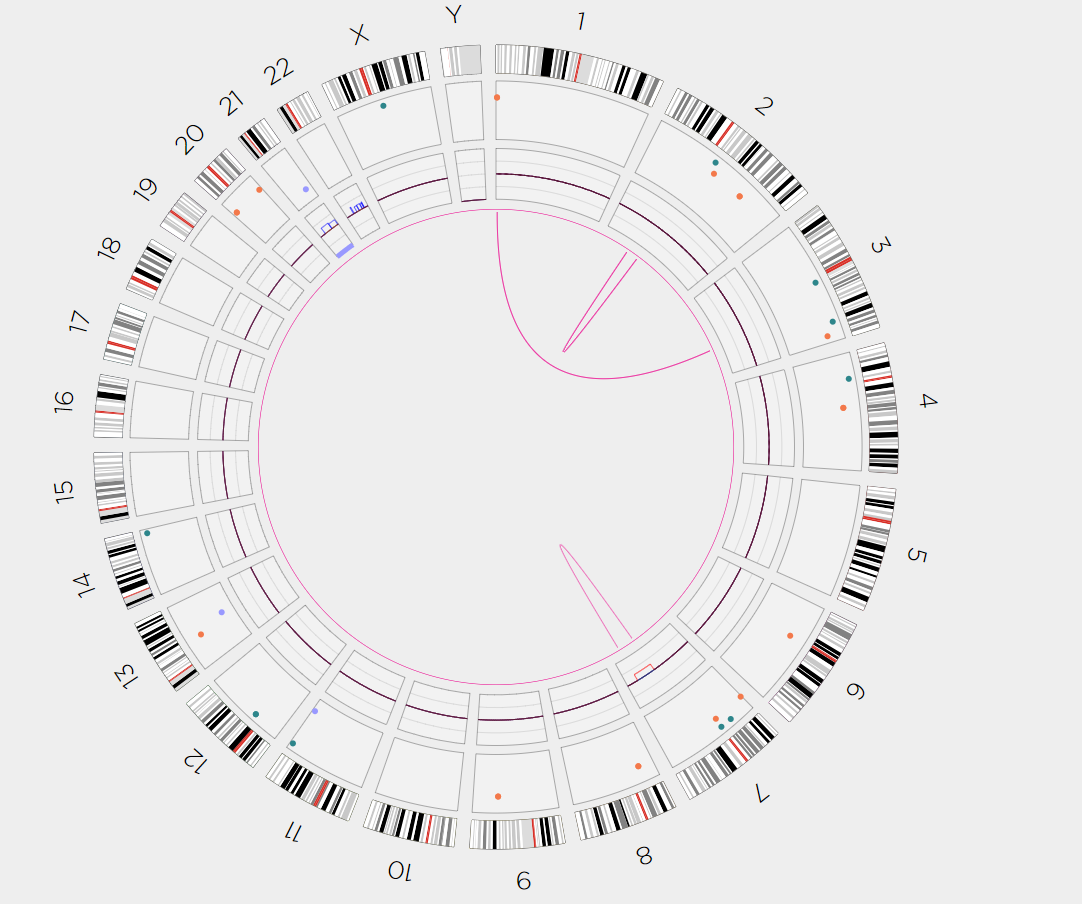
“In our initial clinical samples, we observed some very interesting results,” says Dr. Adam Smith, Director of the Cancer Cytogenetics Laboratory. “The initial individual tested with Optical Genome Mapping showed a unique 3-way translocation, specifically t(3;7;12). This rearrangement was shown to be a recurrent rearrangement involving the MECOM and ETV6 genes – which has an important prognostic significance. The results were reported within a week, without the need for confirmatory FISH testing, which would have been obligatory if a karyotype analysis had been conducted.”

Another sample that came shortly thereafter had a rare recurrent translocation, a t(1;3) –PRDM16::RPN1.
“We would not have been able to confirm this result by conventional cytogenetics as we don’t have a FISH probe that can confirm this rearrangement,” says Dr. Smith.
The t(1;3) is a recurrent translocation that is now included in the latest version of the World Health Organization (WHO) Classification of Hematolymphoid Malignancies (5th Edition).
The UHN Cancer Cytogenetics lab is a national and international leader in the development of Optical Genome Mapping. The detection of chromosomal changes at high resolution will improve treatment selection for patients resulting in better outcomes for Ontarians.
“Congratulations to the Cancer Cytogenetics Laboratory for the development and innovation that this technology brings to UHN, and the resulting improvement in patient care. A truly historic moment for our Cancer Cytogenetics Laboratory,” says Christine Bruce, Senior Director, LMP.

Leave a comment